Updated:
How Does NNMT Help Breast Cancer Cells Spread to the Lungs?
SUMMARY
Metastasis is a major cause of death in patients with solid cancers, but how cancer cells adapt and establish themselves in distant organs is still not fully understood. This study investigated the role of nicotinamide N-methyltransferase (NNMT) in metastatic colonization in basal-like breast cancer. Using cell and mouse models, the researchers showed that NNMT helps maintain a cellular program that supports cancer cell plasticity and lung colonization, partly through the regulation of PRDM5 and COL1A1.
Key Highlights
| Criterion | Description |
|---|---|
| NNMT and patient survival | Patients with strong NNMT expression had a 5-year survival rate of ~43%, vs ~69–73% for low/negative NNMT expression. |
| NNMT and tumor aggressiveness | Strong or intermediate NNMT expression was found in ~25% of 625 primary tumors, and was linked to ERα-negative (more aggressive) tumors. |
| NNMT removal and metastasis | Removing NNMT strongly reduced metastatic colonization in mouse models. |
| COL1A1 restoration | Restoring COL1A1 in NNMT-depleted cells partially restored their ability to colonize the lungs. |
Why do some breast cancer cells metastasize more easily?
Metastasis occurs when cancer cells leave the primary tumor and establish new tumors in other organs — a process that requires them to adapt to new environments called cellular plasticity.
The researchers focused on basal-like breast cancer, an aggressive subtype, and looked for metabolic genes that could drive this adaptability. They identified nicotinamide N-methyltransferase (NNMT), which was strongly linked to aggressive, adverse clinical outcomes.
Removing NNMT reduces metastatic colonization
To test whether NNMT directly contributes to metastasis, the researchers generated knockout breast cancer cells and compared them with cells that still expressed the enzyme. Metastatic progression was tracked over time using bioluminescence imaging, acquired with either an IVIS Spectrum or the Vilber Newton 7.0 system. Results showed a clear reduction of metastasis.
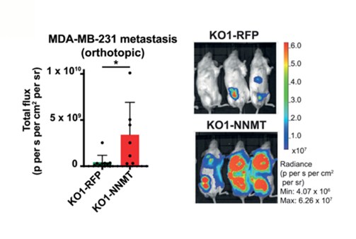
Figure 1. Bioluminescence imaging of mice with breast cancer metastases, using MDA-MB-231 cells in which NNMT had been knocked out (KO). Mice labeled "KO1-RFP" (top) carry NNMT-knockout cells with a control marker (RFP) and show little signal, indicating few metastases. Mice labeled "KO1-NNMT" (bottom) carry the same knockout cells with NNMT reintroduced, and show a much stronger signal — illustrating the increased metastatic colonization driven by NNMT.
How does NNMT affect cancer cell behavior?
The researchers then investigated what happens inside cells when NNMT is removed, and found something counterintuitive. NNMT normally uses methyl groups in a reaction that helps regulate the cell's methylation balance. Without NNMT, these methyl groups accumulate (a "methyl overflow"), and lead to increased DNA and histone methylation at certain genes, switching them off — including PRDM5, a protein that regulates the activity of other genes.
This silencing of PRDM5 was accompanied by a drop in several collagen genes, components of the extracellular matrix, the network of proteins surrounding cells. Among them, COL1A1 stood out as the most consistently affected across all models tested — making it a candidate to explain how NNMT promotes metastasis.
Connecting PRDM5 and COL1A1 to lung colonization
The researchers next tested whether PRDM5 and COL1A1 were actually involved in metastasis, rather than simply being associated with NNMT. Reducing PRDM5 in MDA-MB-231 cells lowered COL1A1 expression and decreased lung metastatic burden. However, restoring COL1A1 in NNMT-depleted cells partially rescued their ability to colonize the lungs, reaching an intermediate level compared with NNMT-expressing cells.
Together, these results identify an NNMT–PRDM5–COL1A1 pathway, in which NNMT sustains PRDM5 activity to drive collagen production, helping breast cancer cells establish themselves in the lungs.
Conclusion
This study reveals a surprising metabolic mechanism behind metastasis: by consuming methyl groups, NNMT maintains an epigenetic state that allows PRDM5 and collagen genes such as COL1A1 to remain active.
Without NNMT, excess methyl groups silence these genes, blocking cancer cells' ability to colonize the lungs — a link the team also confirmed in human tumor samples, where NNMT-positive, hormone receptor-negative tumors showed higher PRDM5 and COL1A1 levels.
Further research will be needed to determine whether this mechanism extends to other cancer types, and whether targeting NNMT could help prevent or slow metastatic spread.
Glossary
Metastasis: Process that happens when cancer cells break off from the original tumor and spread to other areas of your body.
Extracellular matrix: The structure that surrounds cells and provides support and structural organization to tissues.
MDA-MB-231: A human cell line commonly used to model late-stage breast cancer.

Application Specialist & Asia Pacific Manager